
Human Eye Part 1
- By Admin Koushi
- (0) comments
- June 25, 2025
Human Eye Part 1
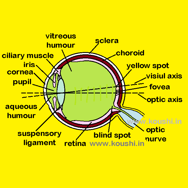
Human eye is spherical in shape with a slight bulge at the front.
Sclera: The outer part of eye is called sclera. It is white in colour with fibrous layer.
The transparent part of it at the front of the outer side of the eye is called cornea. Light enters the eye through it. Its refractive index is about 1.33. Inside of sclera there is a black coloured layer of tissue which is called choroid. It supplies blood to the eye and reduces reflection of light within the eye.
Aqueous humour: It is a transparent liquid of refractive index 1.33. It acts as a refracting medium between cornea and eye lens.

Iris: Iris is a contractile circular ring with a circular hole at its centre called pupil. By adjusting the size of pupil, iris can control the intensity of light entering. Iris closes the pupil to reduce the amount of light entering the eye when a bright object is viewed.
Eye lens: It is a biconvex converging lens made of jelly like transparent material of refractive index about 1.44. It is suspended behind retina by suspensory ligament. Eye lens forms real and inverted image of external object on retina. The focal length of eye lens changes with respect to the position of object.
Ciliary muscle: Suspensory ligaments are attached to a circular ring of muscle is called ciliary muscles. It controls the shape of the lens. When the ciliary muscle is relaxed, the tension in the lens is more. The curvature of lens becomes more hence it can focus the distant object into the retina. Contraction of ciliary muscle makes the lens with less tension. Therefore, the focal length of eye lens decreases which can able to focus the near object at retina.
Vitreous humour: It is a transparent medium of refractive index about 1.33 in between lens and retina. It acts as a refracting medium between cornea and eye lens.
Retina is the inner layer of the eye containing light sensitive cells and nerve fibres. When light falls on the retina, it produces chemical change in the cell which send electrical signals along the nerve fibres through the optical nerve to the brain.
It contents two types of cells called rod and cones. The maximum part of retina contents rod cells which are sensitive to a low level of light but do not give much details or sharpness to the image. There is a portion of diameter about 2 mm at the middle part of retina is called yellow spot. This part is most effective to understand the colour and details of the object. At the centre of yellow spot there is a circular portion of diameter about 0.3 mm is known as fovea. It contains cone cells around fovea which gives the best detailed and coloured vision to the image. So, the muscles of eye always try to cast the image at that region. Millions of nerve fibres leave the retina from a point called blind spot. It contains no light sensitive cells.
Optical axis and visual axis: The line joining centre of the cornea and that of the lens is called optical axis. The line joining centre of lens and fovea is called visual axis. The angle between optical and visual axis ranges from 50 to 70.
Accommodation of eye: It is the ability of eye lens to adjust its focal length to see the objects at all distance.
Persistence of vision: The image formed at retina leaves an impression in our brain for a time span of ![]() second. So, if two different incident occurs before our eye within
second. So, if two different incident occurs before our eye within ![]() second, we cannot differentiate them. We can think that a single incident takes place. For an example, we cannot distinguish the position of a blade of revolving electric fan.
second, we cannot differentiate them. We can think that a single incident takes place. For an example, we cannot distinguish the position of a blade of revolving electric fan.

